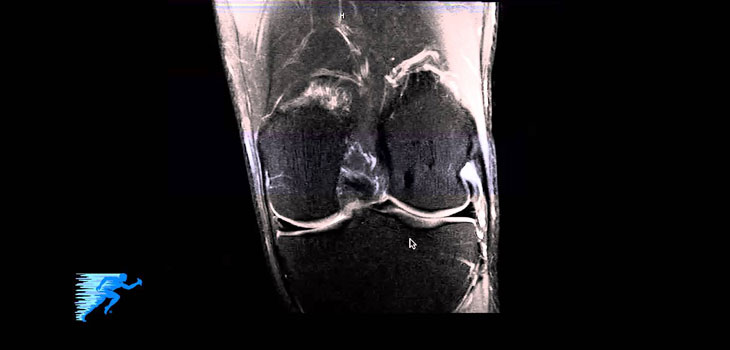
Robert LaPrade, M.D., Ph.D., an orthopedic knee and sports medicine surgeon at The Steadman Clinic, has launched a new video series that is meant to educate non-orthopedic physicians, medical students and patients on reading an MRI of the knee. In this video series, “How to Read a MRI of the Knee, ” Dr. LaPrade reviews individual MRIs and identifies the various anatomic structures and visual indicators of common knee injuries.
Dr. Robert LaPrade Creates Video Series for Doctors, Patients
2 min read Premium comments
Secondary
As indicated in the news release, “Once the healthy anatomical structures are established Dr. LaPrade demonstrates how to read a MRI of the knee for ACL, PCL, MCL and PLC [anterior cruciate ligament, posterior cruciate ligament, medial cruciate ligament, and posterolateral corner] injuries, meniscal root tears, medial meniscus tears and osteochondritis dissecans lesions. In each case, he thoroughly examines all three MRI views to identify if there is bone bruising or an additional injury present.”
Dr. LaPrade told OTW, “I did this project because I believe it is essential that orthopaedic surgeons read their own MRI scans. We know the anatomy, have the benefit of a physical exam to match the anatomy and also need to confirm the findings ourselves.”
“It has been a bit frustrating to me that some of our incoming fellows will ask to look at the MRI report instead of actually viewing the MRI scans themselves. I can understand that sometimes some of them may not have exposure to some of the complex knee injuries that we see, but one should be able to interpret meniscal tears, meniscal root tears, ACL tears and other common pathology. By the end of their year here, I believe all of our fellows have excellent skill levels in reading most knee MRI scans. I found myself teaching the same points over and over again because I can only teach a limited number of sports fellows and Certified Athletic Trainer fellows each year.”
“I thus decided to create this education tool to benefit medical students, residents, fellows and other levels and types of physicians to help understand how to read a knee MRI scan. This may be especially helpful when one needs to interpret an MRI with a patient in the office and a radiology report may not be available for several days. It may also prove useful to those who aspire to be an orthopaedic surgeon and wish to learn more about the knee prior to or during their clinical rotations.”
“To date I have had excellent feedback from physicians and therapists about this educational tool. That was my main goal.”
React:
Discussion
This is a fascinating development. In my practice we've seen similar outcomes with the revised protocol. The key differentiator seems to be patient selection criteria. Has anyone else noticed the correlation with BMI thresholds?
Great point. I'd push back slightly on the conclusion, the sample size in the cited study is too small to draw population-level inferences. That said, the directional signal is compelling and worth a larger RCT.
We implemented a similar approach last year. Early results are promising but we're still gathering 12-month follow-up data. Happy to share our protocol if anyone is interested.
Join the conversation
Orthopedic professionals are discussing this. Sign in and upgrade to read every comment and add your voice.