Breathtaking: World’s First Bilateral Hand Transplant in a Child
Breathtaking: World’s First Bilateral Hand Transplant in a Child// Nonunion Rate in Subtalar Fusions…Mysteries Remain // and More!
6 min read Premium comments
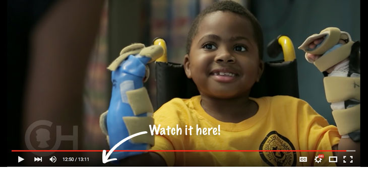
What kind of surgery could involve 18 months of planning, 3 hospitals, 40 medical personnel, and last for 10 hours? A world’s first…a bilateral hand transplant in a child.
The transplant team, led by L. Scott Levin M.D., FACS, chair of orthopaedic surgery at the University of Pennsylvania and professor of surgery (division of plastic surgery), also included medical personnel from the Children’s Hospital of Philadelphia and Shriners Hospital for Children in Philadelphia. Dr. Levin, told OTW, “This brave young boy, Zion Harvey, was referred to us by Scott Kozin and Dan Zlotolow, two talented hand surgeons at Shriners. He had lost his hands at the age of two due to an infection. Now, he has a second chance.”
Dr. Levin is the only U.S. orthopedic surgeon to ever lead a Vascularized Composite Allotransplantation team. He noted, “Zion’s surgery required, among many other individuals, five orthopedic surgeons and five plastic surgeons. The planning was very complex, with involvement required by pharmacy, ICU, nursing, OR, anesthesia, the internal medicine etc.…as well as numerous meetings to explain what was being done. We created extensive checklists and practiced the operation several times at the Penn Human Tissue Laboratory. We utilized CT generated cutting guides for the osteotomies and we used 3D printing to create models of the patient’s hands (based on MRI data) and thus increase efficiency in the OR.”
“We closely followed our protocols and checklists in order to ensure that no step was eliminated, and that what we had rehearsed was exactly what we did during the surgery. Things moved along very efficiently in the OR and we had excellent communication between the entire surgical team. It was a big orchestra. It helped immensely that the technical aspects, such as connecting the donor’s forearm bones to Zion’s arms, were worked out in advance.”
“One sign of success as the operation progressed was that Zion’s new hand was revascularized in the appropriate amount of time. The color and size matching was excellent because we had created models of his ideal hand based on his MRI and CT data. The 3D printing helped us select and confirm that the size of the donor hand was acceptable to transplant onto Zion’s forearm.”
“This has been an unusual, emotional experience…and it must be said that while one little boy has new hands, there is a family that is mourning the loss of their little boy. They deserve the utmost respect.”
“We check in on Zion two to three times a day to ensure that things are progressing as they should. He is already moving his fingers and picking up objects; he will continue with hand therapy for some time.”
“We have made a lifelong commitment to this patient and his family. Because Zion lost his hands at such a young age, we will be doing functional MRI scans of his brain to determine how his brain responds to having hands and how the brain will grow and adapt.”
“In order to do this kind of case you need microvascular experience in free tissue transfer and pediatric microsurgery. Unfortunately, these skills have been lost in American orthopedics; modern microvascular surgery is now predominately the domain of reconstructive plastic surgery. I have been privileged to train a few orthopedic hand and microsurgery fellows who are doing elective reconstructive microsurgery. But this is intensive, and not for everyone. There are not, for example, a lot of joint replacements or arthroscopies that you have to take back in the middle of the night.” This can happen and does happen occasionally in free tissue transfer if there is a vascular thrombosis.
“Zion required urgent return to the operating room due to arterial thrombosis one of his blood vessels. We fixed the problem without incident, but it demonstrates that there is a high degree of vigilance and postop intensity that is required particularly in microsurgery.”
“Zion will be the first of many children to have this surgery; it holds great promise.”
Subtalar Fusions: Digging Into the Nonunion Rate
What is behind the nonunion rate in the subtalar joint? Famed foot and ankle specialist, Chris Coetzee, M.D., wanted to find out. He tells OTW, “I am the primary investigator on a current multicenter study looking at subtalar fusions; we know the nonunion rate is roughly 10% for these procedures, which is pretty high for an orthopedic procedure rate.”
“The fact that the subtalar joint has a fairly poor blood supply may be one thing that contributes to the high nonunion rate. Also, this is a joint with high load sharing…and it’s a small joint, so every time you walk it is absorbing your full body weight. There is also a shear component to load, not only axial”
Dr. Coetzee, noting that this is a prospective randomized controlled trial, added, “To see which treatment would result in a higher fusion rate, in one arm of the study we used stem cells and in the other arm we used autologous bone graft. While we were hoping that the stem cells would be the missing link, it looks like there is no difference between the two groups. Once all the data is collected we will stratify the data to see if there are specific situations were stem cells might be beneficial or superior to autologous graft.”
“We took x-rays at 6 weeks, 3 months, 6 months, and one year; we did CT scans at 6 months. The one thing we learned early on during the study is that the specificity of an x-ray to show fusion is only 30%. The subtalar joint looks like a saddle, so depending on the direction of the x-ray beam things can be unclear. If there is flatfoot involved, then you have to aim the beam perpendicular to the joint in order to determine if it is fused. You really must use a CT scan. While they used to be expensive and a bit challenging to access, you can now have them done just about anywhere—and have them done without a substantial cost.”
If not stem cells, then what is the missing link? Dr. Coetzee says, “It might be that there will just be a certain percentage of subtalar fusions that will result in nonunions. We need to determine if there are different fixation methods that are more effective. At this point we use screws; in theory that is static compression, so if there is a bit of bone resorption then you lose your compression. The idea would be to have something that is ongoing, i.e., dynamic compression such that even if you get bone resorption you will obtain compression across the joint. As for this study, it is ongoing, and we will have the final results by the end of the year.”
Medial Chondrosis Associated with BMI, Alignment, and Medial Meniscal Status
It looks like those patients having to undergo revision ACL [anterior cruciate ligament] reconstruction (rACLR) could be at a disadvantage when it comes to their cartilage. Robert H. Brophy, M.D. is an associate professor of orthopedic surgery at Washington University School of Medicine in St. Louis. Dr. Brophy told OTW, “Patients who undergo rACLR often experience damage to the articular cartilage (chondrosis). We thought that malalignment could overload one compartment of the knee and be associated with a higher rate of chondrosis in that compartment.”
“In one of our previous studies we looked at patients who had undergone a prior meniscus repair and how that related to cartilage wear. We showed that if the patient had had the meniscus removed then they fared worse than if they had undergone a repair. We didn’t know whether it mattered if the repair resulted in a healed meniscus, we were just basing the results on the previous surgery.”
“Using data from the Multicenter ACL Revision Study (MARS) study, a prospective cohort, we were able to identify 246 patients who could be included in the study. We suspected that body mass index (BMI) would be related to chondrosis in the medial compartment; here, the average BMI was 26.4. We found that the medial compartment had more chondrosis than the lateral compartment. Disruption of the meniscus was noted in 35% of patients on the medial side and 16% in the lateral side. Medial compartment chondrosis was associated with BMI, alignment, and medial meniscal status. Lateral compartment chondrosis was significantly associated with age and lateral meniscal status. Patients who had an intact meniscus decreased their odds of having chondrosis by 64% to 84%.”
“While we found no significant association of chondrosis in the lateral compartment with alignment it is possible that the study was underpowered. The study design can show association but not causation, and we’re still talking about a single snapshot of data.”
“We still don’t know if we should correct malalignment in people undergoing ACL reconstruction to prevent arthritis. This needs to be examined prospectively, but this is a challenging study to conduct in adequate numbers.”
React:
Discussion
This is a fascinating development. In my practice we've seen similar outcomes with the revised protocol. The key differentiator seems to be patient selection criteria. Has anyone else noticed the correlation with BMI thresholds?
Great point. I'd push back slightly on the conclusion, the sample size in the cited study is too small to draw population-level inferences. That said, the directional signal is compelling and worth a larger RCT.
We implemented a similar approach last year. Early results are promising but we're still gathering 12-month follow-up data. Happy to share our protocol if anyone is interested.
Join the conversation
Orthopedic professionals are discussing this. Sign in and upgrade to read every comment and add your voice.