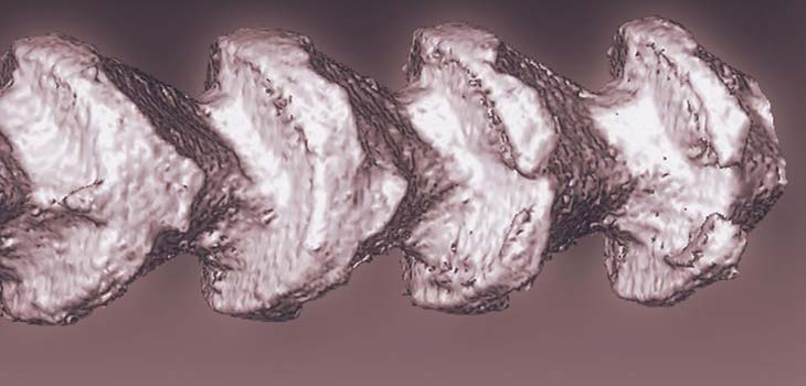
In what may be a unique approach to wound healing, researchers have developed a new approach for better integrating medical devices with biological systems. The researchers, led by Bozhi Tian, Ph.D., assistant professor in chemistry at the University of Chicago, have created a world’s first: skeleton-like silicon spicules prepared via chemical processes.
Silicon for Tissue Regeneration?
2 min read Premium comments
Secondary
“Using bone formation as a guide, the Tian group has developed a synthetic material from silicon that shows potential for improving interaction between soft tissue and hard materials, ” said Joe Akkara in the July 8, 2015 news release. Akkara is a program director in the National Science Foundation materials research division, which funds this research. “This is the power of basic scientific research. The Tian group has created a material that preliminarily seems to enhance soft tissue function.”
According to the news release, Dr. Tian and his colleagues from Northwestern University “achieved three advances in the development of semiconductor and biological materials. One advance was the demonstration, by strictly chemical means, of three-dimensional lithography. Existing lithographic techniques create features over flat surfaces. The laboratory system mimics the natural reaction-diffusion process that leads to symmetry-breaking forms in nature: the grooved and notched form of a bee stinger, for example. Tian’s team developed a pressure modulation synthesis, to promote the growth of silicon nanowires and to induce gold-based patterns in the silicon. Gold acts as silicon’s growth catalyst. By repeatedly increasing and decreasing the pressure on their samples, the researchers were able to control the gold’s precipitation and diffusion along the silicon’s faceted surfaces.”
“One of the major hurdles in the area of bioelectronics or implants is that the interface between the electronic device and the tissue or organ is not robust, ” noted Dr. Tian.
The team indicated that these new spicules may circumvent this issue because they penetrate the collagen and then become deeply rooted.
Tian told OTW, “We developed a new 3D lithography method for preparing complex semiconductor materials. This 3D lithography involves reaction-diffusion cycles which we typically see in the formation of naturally occurring biominerals. The silicon materials we showed in our Science paper exhibit strongest mechanical interactions with extracellular matrix materials-collagen, as compared to other available silicon materials. This property is certainly nice for potential implant applications.”
“The anisotropic, skeleton-like silicon provide a biomimetic mechanism for enhanced interaction with tissues and cells, suggesting their potential as element for bioelectronics (either sensing or stimulation). In particular, we are interested in electroceuticals (i.e., stimulation), in which electrical impulses are delivered through devices to target the neural circuits that regulate the body’s organ functions. Any bioelectronic device works through electrical signal transduction at the bio-interfaces, and the efficacy of such transduction is closely related to the device-bio junction tightness. Our skeleton-like silicon has shown significantly stronger attachment to extracellular matrix materials. We are planning to use these ‘sticky’ silicon as building blocks for making flexible and macroscopic devices capable of peripheral nerve stimulation and wound healing, where electric current/field can be used for controlling cellular physiology and motility.”
“Our skeleton-like materials are made of silicon, which is electrically conductive. One possibility is to use these materials for electrical stimulation to promote tissue regeneration.”
React:
Discussion
This is a fascinating development. In my practice we've seen similar outcomes with the revised protocol. The key differentiator seems to be patient selection criteria. Has anyone else noticed the correlation with BMI thresholds?
Great point. I'd push back slightly on the conclusion, the sample size in the cited study is too small to draw population-level inferences. That said, the directional signal is compelling and worth a larger RCT.
We implemented a similar approach last year. Early results are promising but we're still gathering 12-month follow-up data. Happy to share our protocol if anyone is interested.
Join the conversation
Orthopedic professionals are discussing this. Sign in and upgrade to read every comment and add your voice.