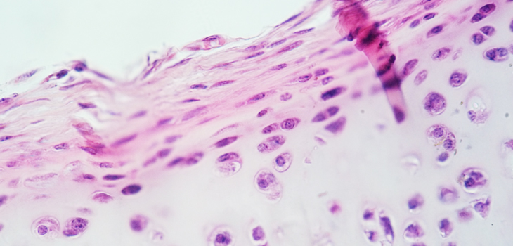
It is an unfortunate fact of life that hyaline cartilage, which cushions joints, does not have the capacity to regenerate. Once it is gone, it is gone forever, according to Professor Norihuki Tsumaki, of the Center for iPS Cell Research and Application at Kyoto University, Japan. Hyaline cartilage consists of chondrocytes and their secretions. The secretions are extracellular matrix (ECM) proteins called collagens II and XI. Significantly, they do not include collagen I, which is the primary collagen that creates fibrocartilage, or scar tissue.
Cartilage Yields Secrets to Japanese Researchers
2 min read Premium comments
Secondary
The key, then, to a successful recovery from a deteriorated joint, according to Tsumaki, is to introduce into the bad joint cartilage chondrocytes that secrete only hyaline cartilage ECM proteins and not collagen I.
One way to do this is via autologous chondrocyte transplantation which involves getting hyaline cartilage from a biopsy and transplanting it into the site of the deteriorated joint. Because the biopsy is smaller than the area being repaired, the chondrocytes must be enlarged. To enlarge the chondrocytes requires the enzymatic digestion of the ECM proteins. The chondrocytes expand, as planned, but in the process they secrete collagen I making the presence of fibrous tissue inevitable after such operations, “The chondrocytes lose their chondrogenic properties, ” laments Tsumaki.
To solve this problem, Tsumaki and his team, in cooperation with a group led by Professor Shuichi Matsuda of KU Graduate School of Medicine, have developed a new protocol that expands not the chondrocytes, but iPS cells. When enough iPS cells are expanded, Tsumaki differentiates them into chondrocytes. Because these chondrocytes are differentiated directly from iPS cells, they have no need to digest ECM proteins, which avoids the problem of fibrous tissue and allows for only hyaline cartilage to be synthesized. Whew!
Because the chondrocytes have already begun secreting ECM proteins, they can be transplanted into joints without using scaffolds. The team has transplanted their cells into three animal models: mouse, rat and mini-pig, and report positive signs for integration and maintenance. Tsumaki said, “These findings are only preliminary, but they show good indications of safety. The next step is to find the best conditions for transplantation in larger animals before we can consider patient treatment.”
React:
Discussion
This is a fascinating development. In my practice we've seen similar outcomes with the revised protocol. The key differentiator seems to be patient selection criteria. Has anyone else noticed the correlation with BMI thresholds?
Great point. I'd push back slightly on the conclusion, the sample size in the cited study is too small to draw population-level inferences. That said, the directional signal is compelling and worth a larger RCT.
We implemented a similar approach last year. Early results are promising but we're still gathering 12-month follow-up data. Happy to share our protocol if anyone is interested.
Join the conversation
Orthopedic professionals are discussing this. Sign in and upgrade to read every comment and add your voice.