MedShape, Inc. has announced that the company has received FDA 510(k) clearance for its FastForward Bone Tether Plate, a product featuring the company’s latest technology platform: the 3D printing of medical grade titanium alloy “that allows for the fabrication of devices with complex and/or customizable geometries.”
MedShape: 510(k) for Bone Tether Plate
2 min read Premium comments
Secondary
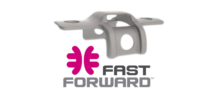
The plate is the main component of the FastForward Bunion Correction System, “a new approach to surgically correct hallux valgus deformities that preserves and protects the native bone anatomy.” According to the February 2, 2015 news release, tethering suture material between the first and second metatarsals is one alternative bone-sparing approach to treatment. “However, peer-reviewed studies have reported the occurrence of second metatarsal fractures due to stress concentrations created by the suture-button implant and the requirement to drill through the second metatarsal.”
“The FastForward Bone Tether Plate represents a breakthrough in bunion correction by allowing suture tape to be securely and safely wrapped around the second metatarsal eliminating the need to drill through the bone. Thanks to the new 3D printing technology, the Bone Tether Plate is equipped with several unique design features including an overall geometry that closely matches the second metatarsal anatomy so that stresses are optimally distributed on the bone and a looped portion that allows suture tape to pass and securely hold the plate in place without the need for bone screws.”
MedShape has also received 510(k) clearance for its FastForward PEEK Screw System, to be used in conjunction with the Bone Tether.
“The FastForward system aligns with MedShape’s continued mission to develop and commercialize innovative medical devices from cutting edge materials and manufacturing technologies to address significant clinical needs, ” said Ken Gall, Ph.D., chief technology officer of MedShape and professor of Materials Science and Engineering at the Georgia Institute of Technology. “We are in the early stages of tracking clinical outcomes with this new surgical approach and are excited to extend the 3D printing material platform towards the development of other implants.”
Dr. Gall told OTW, “In order to address the clinical demands associated with bunion procedures, it was important that we develop a device that would fit securely around the 2nd metatarsal while protecting the bone from large stress risers. By 3D printing the FastForward Bone Tether Plate, we were first able to design the plate with a geometry that aligns closely with the second metatarsal anatomy including side wings that help broadly distributing stresses across the bone. The second feature we were able to incorporate through 3D printing was a low profile loop on the top of the plate that suture tape can pass through to aid in fixating the plate to the bone without the tape coming in contact with bone and creating stress concentrations.”
React:
Discussion
This is a fascinating development. In my practice we've seen similar outcomes with the revised protocol. The key differentiator seems to be patient selection criteria. Has anyone else noticed the correlation with BMI thresholds?
Great point. I'd push back slightly on the conclusion, the sample size in the cited study is too small to draw population-level inferences. That said, the directional signal is compelling and worth a larger RCT.
We implemented a similar approach last year. Early results are promising but we're still gathering 12-month follow-up data. Happy to share our protocol if anyone is interested.
Join the conversation
Orthopedic professionals are discussing this. Sign in and upgrade to read every comment and add your voice.