At Tufts University, a team of researchers is reporting a “world’s first”…the discovery that near-infrared fluorescence can be used to detect osteoarthritis changes over time. Specifically, they successfully tracked the development of osteoarthritis (OA) in mice with a fluorescent molecule “probe” that brightened as the disease progressed. The researchers—from Tufts University School of Medicine (TUSM) and the Sackler School of Graduate Biomedical Sciences at Tufts—indicated that the fluorescent molecule detected cartilage loss in the joint.
Fluorescent Molecule Diagnoses, Monitors OA
2 min read Premium comments
Secondary
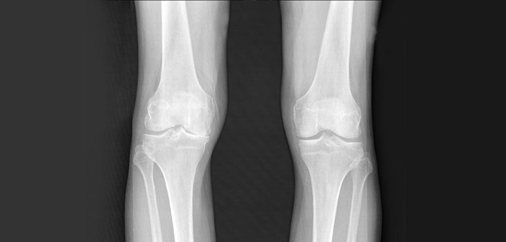
“Patients are frequently in pain by the time osteoarthritis is diagnosed. The imaging tests most frequently used, X-rays, don’t indicate the level of pain or allow us to directly see the amount of cartilage loss, which is a challenge for physicians and patients, ” said co-first author Averi A. Leahy, B.A., an M.D./Ph.D. student in the medical scientist training program at TUSM and the Sackler School, in the February 3, 2015 news release.
“The fluorescent probe made it easy to see the activities that lead to cartilage breakdown in the initial and moderate stages of osteoarthritis, which is needed for early detection and adequate monitoring of the disease. To measure the probe’s signal, we used an optical imaging system, to non-invasively look inside the knee, ” said co-first author Shadi A. Esfahani, M.D., M.P.H., post-doctoral fellow in the division of nuclear medicine and molecular imaging at Massachusetts General Hospital, and in the department of radiology at Harvard Medical School.
One group of mice—54 knees with OA—was given pain medication. The healthy, left knees of the mice served as the control group. Over two months the researchers imaged each knee every two weeks; they found that the probe’s signal became brighter in the injured right knee each time the knee was imaged. The probe emitted a lower signal in the healthy left knee, and did not increase significantly over time.
The corresponding and senior author is Li Zeng, Ph.D., associate professor in the department of integrative physiology and pathobiology at TUSM, and member of the cellular, molecular and developmental biology program faculty at the Sackler School. The work was done in collaboration with Umar Mahmood, M.D., Ph.D., director of the Center for Translational Nuclear Medicine and Molecular Imaging, co-director of Nuclear Medicine and Molecular Imaging, both at Massachusetts General Hospital; and associate professor in the department of radiology at Harvard Medical School.
As a next step the researchers will monitor the fluorescent probe over a longer period of time to determine whether the same results are produced during the late stages of OA.
React:
Discussion
This is a fascinating development. In my practice we've seen similar outcomes with the revised protocol. The key differentiator seems to be patient selection criteria. Has anyone else noticed the correlation with BMI thresholds?
Great point. I'd push back slightly on the conclusion, the sample size in the cited study is too small to draw population-level inferences. That said, the directional signal is compelling and worth a larger RCT.
We implemented a similar approach last year. Early results are promising but we're still gathering 12-month follow-up data. Happy to share our protocol if anyone is interested.
Join the conversation
Orthopedic professionals are discussing this. Sign in and upgrade to read every comment and add your voice.