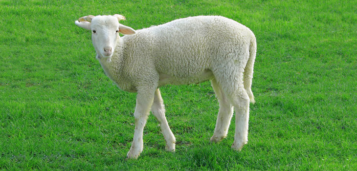
Sheep with regrown meniscus in their knees are happily and painlessly gamboling around their meadow. If the treatment works on sheep—it may on humans as well.
Researchers Grow Meniscus in Sheep’s Knees
2 min read Premium comments
Secondary
Columbia University Medical Center researchers have devised a way to replace the knee’s meniscus by using a personalized 3D printed implant infused with human growth factors to prompt the body to regenerate the meniscus on its own. The procedure, which has been successfully tested on sheep—whose knees are similar to those of humans—could provide the first effective and long-lasting repair of damaged menisci.
Millions of Americans damage their meniscus each year. “At present, there’s little that orthopedists can do to regenerate a torn knee meniscus, ” said study leader Jeremy Mao, DDS, Ph.D., the Edwin S. Robinson Professor of Dentistry (in orthopedic surgery) at the medical center. “Some small tears can be sewn back in place, but larger tears have to be surgically removed. While removal helps reduce pain and swelling, it leaves the knee without the natural shock absorber between the femur and tibia, which greatly increases the risk of arthritis.”
Mao begins his treatment with MRI scans of the intact meniscus in the undamaged knee. The scans are converted into a 3D image. Data from the image are then used to drive a 3D printer, which produces a scaffold in the exact shape of the meniscus, down to a resolution of 10 microns. That is less than the width of a human hair.
The scaffold, which takes about 30 minutes to print, is made of polycaprolactone, a biodegradable polymer. Mao infuses the scaffold with two recombinant human proteins: connective growth factor (CTGF) and transforming growth factor β3 (TGFβ3). Mao’s team found that sequential delivery of these two proteins attracts existing stem cells from the body and induces them to form meniscal tissue.
For a meniscus to properly form, however, the proteins must be released in specific areas of the scaffold in a specific order. This is accomplished by encapsulating the proteins in two types of slow-dissolving polymeric microspheres, first releasing CTGF (to stimulate production of the outer meniscus) and then TGFβ3 (to stimulate production of the inner meniscus).
Finally, he inserts the protein-infused scaffold into the knee. In sheep, the meniscus regenerates in four to six weeks. Eventually, the scaffold dissolves and is eliminated by the body.
“This is a departure from classic tissue engineering, in which stems cells are harvested from the body, manipulated in the laboratory, and then returned to the patient—an approach that has met with limited success, ” said Mao. “In contrast, we’re jumpstarting the process within the body, using factors that promote endogenous stem cells for tissue regeneration.”
“This research, although preliminary, demonstrates the potential for an innovative approach to meniscus regeneration, ” said Scott Rodeo, M.D., sports medicine orthopedic surgeon and researcher at Hospital for Special Surgery in New York City. “This would potentially be applicable to the many patients who undergo meniscus removal each year.”
React:
Discussion
This is a fascinating development. In my practice we've seen similar outcomes with the revised protocol. The key differentiator seems to be patient selection criteria. Has anyone else noticed the correlation with BMI thresholds?
Great point. I'd push back slightly on the conclusion, the sample size in the cited study is too small to draw population-level inferences. That said, the directional signal is compelling and worth a larger RCT.
We implemented a similar approach last year. Early results are promising but we're still gathering 12-month follow-up data. Happy to share our protocol if anyone is interested.
Join the conversation
Orthopedic professionals are discussing this. Sign in and upgrade to read every comment and add your voice.