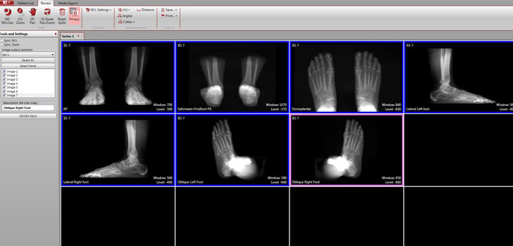
One surgeon, one foot, one scan…the pedCAT system just may be eliminating the need to take multiple X-rays. CurveBeam, LLC is announcing that a new software feature automatically generates unlimited standard x-ray views. The company’s weight bearing CT imaging system now makes it possible for an orthopedic surgeon to capture standard, three-dimensional volume and multi-planar slices in only one click.
CurveBeam: Eliminating Plain X-rays for Foot, Ankle
2 min read Premium comments
Secondary
According to the November 12, 2014 news release, because the x-ray views are computed from the pedCAT 3D data, they are anatomically accurate. Plain radiographs are sensitive to the beam angle of the x-ray tube, and could be distorted or magnified. The company indicates that the pedCAT is the first and only cone beam CT imaging system dedicated to the foot and ankle extremity. pedCAT scans are ultra-low dose, according to a study by Dr. John Ludlow published in the International Journal of Diagnostic Imaging. One scan exposes the patient to the same radiation as a plain film series of the foot and ankle of about three to seven images. The pedCAT’s field of view can capture both entire feet and the bases of the tibia and fibula.
“The software now lets you produce an infinite number of x-ray views from a single pedCAT scan, meaning the patient doesn’t have to be repositioned every time you want to capture a different angle, ”stated Liz Qualtier, CurveBeam vice president of Customer Care. “A pedCAT scan takes about one minute. There is tremendous potential for workflow efficiencies if a pedCAT is used in place of a traditional x-ray unit.”
“Say the physician is looking at a lateral x-ray view and is unsure about whether what he is seeing is a calcaneal fracture, he can quickly switch over to the multiplanar view in CubeVue and scroll through the calcaneus slice by slice for a thorough evaluation, ” Qualtier said. “And he can accomplish all of this within the patient’s initial visit to his office.”
CurveBeam President & CEO Arun Singh told OTW, “The X-ray feature makes the learning curve for pedCAT technology more accessible to all foot and ankle specialists. The leap from plain radiographs to in-office CT imaging can seem daunting. And perhaps surgeons may have thought the pedCAT is similar to its traditional CT counterpart, and indicated for only the most complex cases. We are confident this new feature will cement that the pedCAT has utility for routine pathologies as well.”
OrthoCarolina and the Institute for Foot and Ankle Reconstruction at Mercy are two institutions currently using pedCAT. Singh said that he projects the technology to become commonplace in orthopedic clinics with a devoted foot and ankle section over the next two years.
React:
Discussion
This is a fascinating development. In my practice we've seen similar outcomes with the revised protocol. The key differentiator seems to be patient selection criteria. Has anyone else noticed the correlation with BMI thresholds?
Great point. I'd push back slightly on the conclusion, the sample size in the cited study is too small to draw population-level inferences. That said, the directional signal is compelling and worth a larger RCT.
We implemented a similar approach last year. Early results are promising but we're still gathering 12-month follow-up data. Happy to share our protocol if anyone is interested.
Join the conversation
Orthopedic professionals are discussing this. Sign in and upgrade to read every comment and add your voice.