New work from the University of California (UC), San Diego, may help clarify things when it comes to loss of muscle mass in the elderly. The researchers, led by J.S. Chen, the William Prager Professor of structural engineering at the Jacobs School of Engineering at UC San Diego, are touting the first ever comprehensive numerical simulation of skeletal muscle tissue using a method that uses the pixels in an image as data points for the computer simulation. It is known as mesh-free simulation.
Engineers Simulate Skeletal Muscle Tissue With Pixels
2 min read Premium comments
Secondary
Another area of application for this framework would be the simulation of tissue injuries caused by extreme events such as blasts, car crashes and sport collisions, said Dr. Chen in the September 30, 2014 news release. “This will require adding the mechanics of tissue damage to the simulation model, including how tissue behaves and functions under high velocity impact.”
Dr. Chen’s research group developed their own simulation techniques in order to assess how much force is generated from various muscle systems, and how loss of force relates to loss of muscle volume due to aging. “They found that the loss of force is greater than the loss of volume, which is consistent with prior physiological studies.”
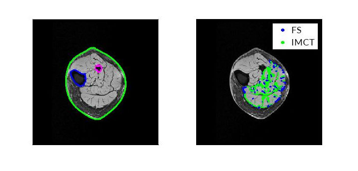
Dr. Chen told OTW, “We developed a new ‘mesh-free’ numerical modeling technique for the modeling of skeletal muscles using data directly from medical image scans such as MRI and CT scans. Using this new computer simulation technique, our initial efforts were to differentiate the causes for loss in force production in the elderly people due to different material composition of the muscle. A few decades ago this loss was mainly attributed to the loss in muscle mass due to aging, termed as ‘sarcopenia, ’ which decreased the force production capacity in the elderly. In more recent research work, it has been observed in many studies that the loss in force due to aging is not only due to the loss of muscle mass but other factors like non-contractile tissue properties, changes in fiber type composition, neural changes, etc., which also contribute to force loss due to aging. Our mesh-free based numerical study investigation found that the loss of force is greater than the loss of volume, which is consistent with prior physiological studies.”
“Modeling can be applied to study other general medical conditions of muscle tissues for example, muscular dystrophy, arthritis, muscular atrophy etc., as well as other biological materials and systems. The results from numerical simulation can provide more information for the doctors and surgeons what are the primary contributors to the disease condition, how much they effect the progression of the disease and help them find a better remedy for the conditions.”
React:
Discussion
This is a fascinating development. In my practice we've seen similar outcomes with the revised protocol. The key differentiator seems to be patient selection criteria. Has anyone else noticed the correlation with BMI thresholds?
Great point. I'd push back slightly on the conclusion, the sample size in the cited study is too small to draw population-level inferences. That said, the directional signal is compelling and worth a larger RCT.
We implemented a similar approach last year. Early results are promising but we're still gathering 12-month follow-up data. Happy to share our protocol if anyone is interested.
Join the conversation
Orthopedic professionals are discussing this. Sign in and upgrade to read every comment and add your voice.