Two physicians from OrthoIndy, neither of whom has any financial or other ties with the manufacturer of this device, have been using a “zipper” instead of sutures for a while and wanted to write about their experiences. After reading their article, we realized that this is one of the coolest new orthopedic technologies in a while and to share it with OTW’s readers. We think you’ll find the idea of zipping instead of suturing mighty interesting. — RY
Zippers Instead of Sutures? This is Cool.
6 min read Premium comments
Companies, academic institutions and their respective R&D departments have historically concentrated their R&D efforts on improving prosthetic joint designs, materials or to improve surgical techniques all with the primary aim of improving outcomes and functional joint longevity.
Based on our experience, however, we would suggest that less “invasive” changes to surgical procedure be considered by physicians particularly if such changes further improve outcomes and overall patient satisfaction.
Zip Surgical Skin Closure
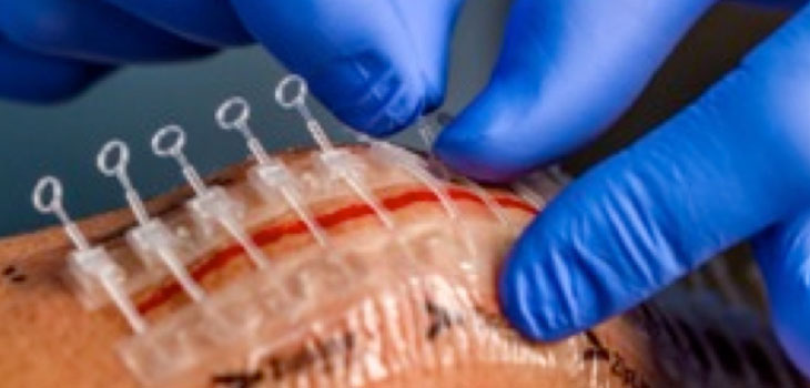
At OrthoIndy, we have been using a new device for closing the skin layer on our knee restoration, UKA/TKA and THA [unicompartmental knee arthroplasty/total knee arthroplasty, and total hip arthroplasty] procedures called “Zip” Surgical Skin Closure (ZipLine Medical, Inc., Campbell, California). When we use the “Zip” Surgical Skin Closure we have observed faster closure time, excellent outcomes as well as improved patient comfort and satisfaction compared to traditional suture and staple closure methods.
A number of published papers (see reference list at the end of this article) have suggested that the device’s non-invasive format may reduce the risk of infection. We also note that several studies on previous tape-based wound closure have also suggested that infection rates are lower compared to staples and sutures.1-5
" data-large-file="https://i0.wp.com/ryortho.com/wp-content/uploads/2014/08/Zippers_ZipSurgicalSkinClosure_WEB.jpg?fit=250%2C298&ssl=1" src="https://i0.wp.com/ryortho.com/wp-content/uploads/2014/08/Zippers_ZipSurgicalSkinClosure_WEB.jpg?resize=250%2C298&ssl=1" alt="Figure 1: Zip Surgical Skin Closure" height="298" width="250">Figure 1: Zip Surgical Skin Closure
When the company’s representatives first presented us with the Zip device, we were intrigued by the concept of being able to close the wound without having to perforate or drag a suture through the skin, which theoretically could be a portal for bacteria to enter.
The Zip attaches to the skin adjacent to the incision using a tenacious hydrocolloid adhesive and utilizes adjustable “zip-tie” straps for wound tensioning. In previous studies, researchers had suggested that closing a wound with an adhesive tape-based closure distributed stress more uniformly to the collagen fibers that cross the wound which, in turn, caused rapid fiber orientation and increased tensile strength.1-6
In our experience, applying the Zip took about the same amount of time as applying staples.
The device’s structure has the potential to distribute closing forces more evenly along the incision compared to staples and subcuticular sutures. We found the wound closure using ZIP to be uniform without being too tight—which, in our view, reduces potential harm to the skin. We found that the device was easy to apply and that we could not only fix the tension, but if necessary, readjust tension later.
With the ZIP closure system, the wound seems to be protected from patient-induced distraction forces during healing. The device has a “programmed separation” which allows it to lengthen during knee flexion without stressing the incision closure.
Typical wear time is 14 days. In our experience, the device was easily and atraumatically peeled from the skin like a dressing to remove.
Some of our patients have been able to remove the device themselves while at home, eliminating the return visit required for the often painful staple removal. In general our patients have found the device to be much more comfortable and less anxiety-inducing compared to staple removal.
Here are the steps we follow to apply the Zip:
- Incision– We apply the Zip device after applying subcutaneous sutures and achieving about a 5mm or smaller incision gap. We have found that good sub-q suture alignment makes the application and closure of the Zip easier.
- Skin Prep– We pull the incise barrier back from the incision and then wipe the skin dry.
- Application– We center the device on the incision, and usually use one complete 16cm Zip for a UKA or TKA, and cut the device shorter for THA. For longer incisions, an additional device may be applied.
- Skin Closure– We tighten each tensioning strap sequentially, starting on one end of the incision, and then go back and adjust individual straps as needed. Although we have not needed to use this, each strap can be released and re-engaged if it is over-tightened. We then cut the strap ends to prevent tampering or catching on dressing or clothing. We usually achieve slight eversion of the incision.
- Dressing- The incision is exposed in the center of the device, enabling an absorptive dressing placed on top to collect wound exudate. We use an absorptive pad over the device, taking care not to allow the dressing adhesive to contact the device.
- Removal- Our patients usually come back for follow-up and Zip removal at POD 14. It is peeled off like a dressing.
" data-large-file="https://i0.wp.com/ryortho.com/wp-content/uploads/2014/08/Zippers_LEAD_SurgicalSkinClosure_WEB1.jpg?fit=730%2C350&ssl=1" src="https://i0.wp.com/ryortho.com/wp-content/uploads/2014/08/Zippers_LEAD_SurgicalSkinClosure_WEB1.jpg?resize=595%2C285&ssl=1" alt="Figure 2: Incision is closed by sequentially tightening straps. / Source: Authors and OrthoIndy" height="285" width="595">Figure 2: Incision is closed by sequentially tightening straps. / Source: Authors and OrthoIndy
Surgeon as Patient
Dr. Farr has had the unique experience of using the Zip for his knee patients and having it used on himself. Dr. Fisher, co-author of this article, performed a UKA on Dr. Farr and closed the wound with the ZIP device. Four days following surgery and with the ZIP on his knee, Dr. Farr was using a stationary bicycle. The Zip’s Dynamic Compression feature maintained good wound closure while not impeding his range of motion. The closure device was able to lengthen with the incision during knee flexion. Dr. Farr found the device to be both comfortable and painless to remove, with a good cosmetic result—just like his own patients.
Reducing Patient Apprehension
We found the device helped to reduce patient apprehension, which is frequently associated with staple removal. This improved the staff-patient interaction and contributed to greater patient satisfaction.
We remove the device at a slightly longer time-frame postoperatively (POD 14) than staples (POD 10) because we do not have the concern of erythema or “cross-hash marks” that may occur with longer staple retention. ZIP is removed atraumatically by simply peeling it off the skin, as with a dressing. That, as much as anything, served to eliminate our patient’s apprehension and pain which is more commonly associated with staple removal.
We have had high acceptance with our patients and they are quite happy with the appearance of their scar after the device is removed.
Our initial experience has been positive and we are integrating the Zip into our clinical practice.
In our collective, long experience we started with sutures and then evolved to staples. The next evolution, at least in our practice, is to this non-invasive Zip device. As with any new device, further studies will be needed to further quantify the benefits, and we are planning a randomized, controlled prospective study on bilateral partial or total knee patients to measure the differences between the Zip and staples for total and partial joint procedure skin closure.
_______________________________________________________
1 Smith TO, Sexton D, Mann C, Donell S. Sutures versus staples for skin closure in orthopaedic surgery: meta-analysis. BMJ. 2010 Mar 16;340:c1199
2 Schauerhamer RA, et al. Studies in the Management of the Contaminated Wound, VII. Susceptibility of Surgical Wounds to Postoperative Surface Contamination. Am J Surg 1971; 122:74-77.
3Henretig FM, King C. Textbook of Pediatric Emergency Procedures, Chapter 111 Laceration Repair, p. 1159.
4Hirshman HP, Schurman DJ, Kajiyama G. Penetration of Staphylococcus aureus into Sutured Wounds. J Orthop Res, Vol 2, No. 3, 1984.
5Maharaj D, Sharma D, Ramdass M, Naraynsingh V. Closure of traumatic wounds without cleaning and suturing. Postgrad Med J 2002; 78: 281-282.
6Forrester JC, Zederfeldt BH, Hayes TL, Hunt TK. Wolff’s law in relation to the healing skin wound. J Trauma, 1970; Vol 10, No. 9: 770-779.
React:
Discussion
This is a fascinating development. In my practice we've seen similar outcomes with the revised protocol. The key differentiator seems to be patient selection criteria. Has anyone else noticed the correlation with BMI thresholds?
Great point. I'd push back slightly on the conclusion, the sample size in the cited study is too small to draw population-level inferences. That said, the directional signal is compelling and worth a larger RCT.
We implemented a similar approach last year. Early results are promising but we're still gathering 12-month follow-up data. Happy to share our protocol if anyone is interested.
Join the conversation
Orthopedic professionals are discussing this. Sign in and upgrade to read every comment and add your voice.