The chemical engineers at MIT were out to create a replacement for bone. The current standard for treating bone injuries is to remove a piece of bone from another part of the body and transplant it into the site of the injury. This painful process does not always work well. The MIT engineers set out to do better.
MIT Engineers Marry Polymers with Growth Factors
2 min read Premium comments
Secondary
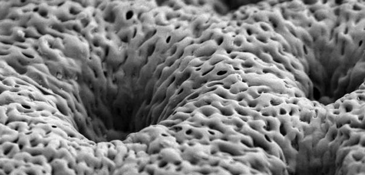
The first step in creating new bone, they realized, was to make a scaffold coated with bone growth factors. They made their scaffold from a polymer called PLGA that is widely used in medical treatment and is biodegradable. Researchers can time it to disintegrate at a specific rate.
The coatings that they put on the scaffold—platelet-derived growth factor (PDGF) and bone morphogenetic protein 2 (BMP-2)—were known to lure the body into producing new bone that is as strong as the original tissue. As Anne Trafto, of the MIT News Office, explained, PDGF is one of the first factors the body releases as part of the wound-healing process following a bone injury. Once PDGF is on the scene other factors, including BMP-2, come in to create the right environment for bone regeneration. Among their other tasks, they recruit cells that can produce bone and blood vessels.
Problem solved, they thought. But it was not. The MIT engineers discovered that when very large quantities of growth factors are delivered too quickly, the body perversely clears them away from the treatment site. Not only do they have a reduced impact on tissue repair, they can also cause unwanted side effects.
Trafto quoted Paula Hammond, the David H. Koch Professor in Engineering and a member of MIT’s Koch Institute for Integrative Cancer Research, as saying, “You want the growth factor to be released very slowly and with nanogram or microgram quantities, not milligram quantities. You want to recruit these native adult stem cells we have in our bone marrow to go to the site of injury and then generate bone around the scaffold, and you want to generate a vascular system to go with it.”
The researchers learned that the growth factors had to be released over a period of several days or even weeks. To slow down the delivery, they devised some ultra-thin porous scaffold sheets that they coated with 40 layers of BMP-2 and, on top of that, another 40 layers of PDGF. “This is a major advantage for tissue engineering for bones because the release of the signaling proteins has to be slow and it has to be scheduled, ” says Nicholas Kotov, a professor of chemical engineering at the University of Michigan who was not part of the research team.
Tests on a rat’s skull were positive. “Using this combination allows us to not only have accelerated proliferation first, but also facilitates laying down some vascular tissue, which provides a route for both the stem cells and the precursor osteoblasts and other players to get in and do their jobs. You end up with a very uniform healed system, ” Hammond said.
Trafton reports that Hammond’s team has filed a patent based on this work and aims to begin testing the system in larger animals with the intent of eventually moving it into clinical trials.
The National Institute of Health funded the project.
React:
Discussion
This is a fascinating development. In my practice we've seen similar outcomes with the revised protocol. The key differentiator seems to be patient selection criteria. Has anyone else noticed the correlation with BMI thresholds?
Great point. I'd push back slightly on the conclusion, the sample size in the cited study is too small to draw population-level inferences. That said, the directional signal is compelling and worth a larger RCT.
We implemented a similar approach last year. Early results are promising but we're still gathering 12-month follow-up data. Happy to share our protocol if anyone is interested.
Join the conversation
Orthopedic professionals are discussing this. Sign in and upgrade to read every comment and add your voice.