The advanced imaging experts at CurveBeam, LLC are announcing the results of a study indicating that the amount of radiation a patient is exposed to during a weight bearing cone beam CT (CBCT) scan is negligible. The research, published in the International Journal of Diagnostic Imaging (Vol. 1, No. 2), found that the radiation dose of a cone beam CT scan of the foot and ankle is comparable to a series of conventional plain radiographs or a few hours of daily background radiation. “Therefore, the dose of radiation should not be a concern when considering the use of cone beam CT imaging for foot/ankle examination, ” the authors said.
CurveBeam: Study Shows Weight Bearing CT Dose is “Negligible”
2 min read Premium comments
Secondary
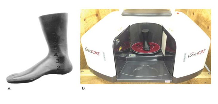
The study was authored by Professor John B. Ludlow, of the Department of Diagnostic Sciences at the University of North Carolina at Chapel Hill. He compared the effective doses of lateral, oblique and AP radiographs with medical CT and cone beam CT images of the foot and ankle. He acquired the cone beam CT images with a pedCAT, the product developed by CurveBeam.
Bony pathologies resulting from trauma and osteoarthritic changes in the foot and ankle are difficult to visualize on plain film imaging, according to the study titled, “Weight bearing CBCT, MDCT [multi-detector computed tomography], and 2D imaging dosimetry of the foot and ankle.”
Medical CT is a diagnostic alternative, however examinations can be expensive and availability is limited to hospitals and large radiology practices. Medical CT scans are also associated with significantly increased X-Ray dose in relation to plain views.
“An additional virtue of CBCT is its high resolution, which permits visualization of fine bone detail and early pathologic changes, ” Dr. Ludlow told OTW.
“This resolution is as good or better than typical MDCT scans, ” Dr. Ludlow said. “While we did not test image quality parameters in our dosimetry study, our sample images had noticeably better resolution than the state of the art MDCT scanner that we used for dosimetric comparison.”
According to the company, the pedCAT is one of the first cone beam CT devices designed for orthopedic specialists. It can image one or both feet while the patient is fully weight bearing. Scans take about 20 to 60 seconds and the complete diagnostic information is available within minutes. Because of its compact size and minimal shielding requirements, the pedCAT is ideal for in-office use.
“Our experience testing CBCT units for dedicated dental and maxillofacial imaging had prepared us to expect doses that were a fraction of MDCT doses, but I was surprised at just how small the dose for foot and ankle imaging was, ” Dr. Ludlow added. “The pedCAT unit generated a range of effective doses that were on the order of a few hours to a maximum of a single day of whole body dose that an adult might receive from ubiquitous background radiation.”
React:
Discussion
This is a fascinating development. In my practice we've seen similar outcomes with the revised protocol. The key differentiator seems to be patient selection criteria. Has anyone else noticed the correlation with BMI thresholds?
Great point. I'd push back slightly on the conclusion, the sample size in the cited study is too small to draw population-level inferences. That said, the directional signal is compelling and worth a larger RCT.
We implemented a similar approach last year. Early results are promising but we're still gathering 12-month follow-up data. Happy to share our protocol if anyone is interested.
Join the conversation
Orthopedic professionals are discussing this. Sign in and upgrade to read every comment and add your voice.