Intravenous iron can be used to effectively label stem cell transplants so they can be tracked in target tissues—such as arthritic joints—according to a recent study conducted at Stanford University School of Medicine.
Iron-Laced Stem Cells
2 min read Premium comments
Secondary
According to the study, intravenous iron can be utilized to effectively label stem cell transplants so they can be tracked in target tissues using magnetic resonance imaging (MRI). As is widely understood, mesenchymal stem cells (MSCs) derived from bone marrow appear to have remarkable potential in the field of cell-based therapy and tissue regeneration.
Heike Daldrup-Link, M.D., associate professor at Stanford University School of Medicine, notes that investigators have long worked with transplanted MSCs to get them to repair damaged joints by promoting the generation of bone, cartilage and connective tissue.
The problem, as Daldrup-Link sees it is that when stem cells are transplanted they often die and disappear from the transplant site. To confirm that a transplant has been successful, the investigator must track the stem cells they have introduced into the body and find out where they are and what they are doing.
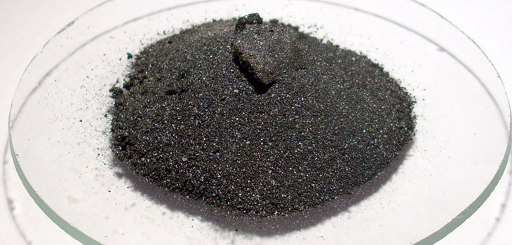
Present practice is to remove the stem cells from the donor and culture them with an iron oxide solution before transplanting them in the patient. This raises the risk of contamination of the stem cells. The Stanford researchers propose that the stem cells can be treated in vivo by giving the donor an iron supplement intravenously prior to the harvesting of the stem cells. Iron oxide, which can be tracked using magnetic resonance imaging (MRI), would be taken up by the donor cells including the stem cells that are due to be transplanted.
Researchers at Stanford University School of Medicine have tried this on rats and it worked. They injected the rats with ferumoxytol, an iron supplement approved for human use, 48 hours before extracting stem cells from their bone marrow. The uptake of iron by stem cells was found to be greater after administration of in vivo ferumoxytol than it had been with the conventional ex vivo-labeling technique.
They transplanted the stem cells into knee cartilage defects of seven rats and tracked them with MRIs for up to a month. Microscopic examination revealed the presence of iron in the stem cells and proved that repair was ongoing in the damaged joints.
This non-invasive monitoring of the accumulation and movement of stem cells, if further developed, will be helpful for clinicians to determine whether cell delivery has taken place in the targeted site.
React:
Discussion
This is a fascinating development. In my practice we've seen similar outcomes with the revised protocol. The key differentiator seems to be patient selection criteria. Has anyone else noticed the correlation with BMI thresholds?
Great point. I'd push back slightly on the conclusion, the sample size in the cited study is too small to draw population-level inferences. That said, the directional signal is compelling and worth a larger RCT.
We implemented a similar approach last year. Early results are promising but we're still gathering 12-month follow-up data. Happy to share our protocol if anyone is interested.
Join the conversation
Orthopedic professionals are discussing this. Sign in and upgrade to read every comment and add your voice.