What are sonic tweezers? From Nanowerk News, an IT publication in the United Kingdom, comes a description that says that sonic/electronic tweezers are gravity-beating ultrasound beams that can grip and hold tiny clusters of cells. Cells in the grip of such devices can be levitated for weeks in a nutrient-rich fluid and be stimulated to grow and form better implant tissue than if the tissue were cultured in a glass petro dish.
Astonishing Sonic Tweezers Manipulate Cells Into Cartilage
2 min read Premium comments
Secondary
By holding the cells in the required position the tweezers can mould growing cartilage tissue into exactly the shape researchers want so that an implant will be the right size and shape when it is inserted into the patient’s knee.
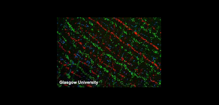
Ultrasonic tweezers have been developed by a closely integrated team of researchers from four United Kingdom universities (Bristol, Dundee, Glasgow and Southampton) together with a select group of industrial partners. Their work has established the UK as a world leader in this technology.
Professor Bruce Drinkwater, of Bristol University who coordinated the program, said, “Ultrasonic tweezers have all kinds of possible uses in bioscience, nanotechnology and more widely across industry. They offer big advantages over optical tweezers that rely on light waves and also over electromagnetic methods of cell manipulation. For example, they have a complete absence of moving parts and can manipulate not just one or two cells (or other objects) at a time but clusters of several centimeters across—a scale that makes them very suitable for applications like tissue engineering.”
Professor Martyn Hill from the University of Southampton, who led the cartilage tissue engineering work, said, “Ultrasonic tweezers can provide what is, in effect, a zero-gravity environment perfect for optimizing cell growth. As well as levitating cells, the tweezers can make sure that the cell agglomerates maintain a flat shape ideal for nutrient absorption. They can even gently massage the agglomerates in a way that encourages cartilage tissue formation.”
The research program has also shown that ultrasonic tweezers can be used to build up cell tissue layer by layer. Members of the team anticipate that the tweezers could be used to help to reconstruct nerve tissue after severe trauma such as limb amputation.
React:
Discussion
This is a fascinating development. In my practice we've seen similar outcomes with the revised protocol. The key differentiator seems to be patient selection criteria. Has anyone else noticed the correlation with BMI thresholds?
Great point. I'd push back slightly on the conclusion, the sample size in the cited study is too small to draw population-level inferences. That said, the directional signal is compelling and worth a larger RCT.
We implemented a similar approach last year. Early results are promising but we're still gathering 12-month follow-up data. Happy to share our protocol if anyone is interested.
Join the conversation
Orthopedic professionals are discussing this. Sign in and upgrade to read every comment and add your voice.