The journal Stem Cell Reports has published the first study to identify the origin cells of human articular cartilage and track their early development. This research could provide a new cell source and biological roadmap for therapies to repair cartilage defects and damage from osteoarthritis, writes Shaun Mason, of Domain-b.
Stem Cell Cartilage Rejuvenation Inches Closer
2 min read Premium comments
Secondary
While scientists have studied the ability of different cell types to generate articular cartilage, up to the present, despite hype, none of the current cell-based repair strategies have worked. None have generated long-lasting articular cartilage tissue in the laboratory. These include exhaustive testing of expanded articular chondrocytes or mesenchymal stromal cells from adult bone marrow, adipose tissue, sinovium and amniotic fluid.
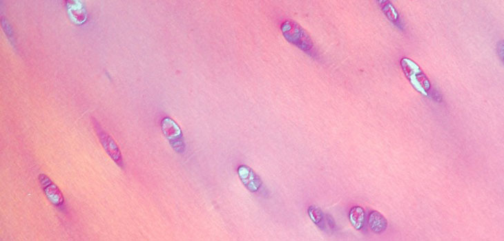
Denis Evseenko, M.D., an assistant professor of orthopedic surgery and head of UCLA’s Laboratory of Connective Tissue Regeneration has now tested and confirmed the growth of progenitor cells into cartilage cells and monitored their growth progress, recording important genetic features that indicate the growth stages of these cells as they develop into cartilage cells.
The researchers used complex molecular biology techniques to determine which cells grown from embryonic stem cells were the progenitors of cartilage cells, or chondrocytes. Mason wrote that, by bridging developmental biology and tissue engineering, Evseenko’s discoveries represent a critical “missing link, ” providing scientists with checkpoints to tell if the cartilage cells are developing correctly.
“We began with three questions about cartilage development, ” Evseenko said. “We wanted to know the key molecular mechanisms, the key cell populations and the developmental stages in humans. We carefully studied how the chondrocytes developed, watching not only their genes but other biological markers that will allow us to apply the system for the improvement of current stem cell–based therapeutic approaches.”
Evseenko’s research was the first to employ animal-free standards to generate the key landmarks that allow the development of cell types that could be used in treatments to re-grow damaged human cartilage. Animal-based components, while they do help stem cells to grow, can lead to variations and contamination, Mason noted. With the goal of producing stem cell serums that are safe for humans, Evseenho could not rely on any animal components.
With the progenitor cells and the landmarks of proper cartilage development identified, Evseenko believes that an effective cellular therapy for diseased or damaged joint cartilage could be tested in human trials within three years.
Mason noted that such stem cell-based therapies would make many current knee and hip replacement surgeries unnecessary. They would offer patients the ability to regrow lost cartilage, keep their bones intact and avoid the risk of major joint-replacement surgery.
React:
Discussion
This is a fascinating development. In my practice we've seen similar outcomes with the revised protocol. The key differentiator seems to be patient selection criteria. Has anyone else noticed the correlation with BMI thresholds?
Great point. I'd push back slightly on the conclusion, the sample size in the cited study is too small to draw population-level inferences. That said, the directional signal is compelling and worth a larger RCT.
We implemented a similar approach last year. Early results are promising but we're still gathering 12-month follow-up data. Happy to share our protocol if anyone is interested.
Join the conversation
Orthopedic professionals are discussing this. Sign in and upgrade to read every comment and add your voice.