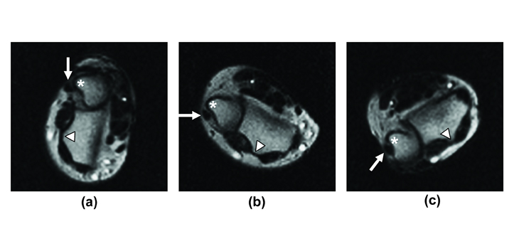
If a still picture won’t work, try a movie. Orthopedic surgeons at the University of California have learned to create “movies” of the wrist in motion—images that are useful in diagnosing the subtle changes in physiology that indicate the onset of conditions such as instability of the wrist.
Moving MRI Useful in Diagnosis
1 min read Premium comments
Secondary
The films are made by creating a series of brief magnetic resonance imaging scans—a technique they call an “active MRI.”
Philip Briggs, who reported on the work from UK’s Spire Gatwick Park Hospital, quoted Robert Boutin, M.D., lead author of the study, who said: “Now patients can reproduce the motion that’s bothering them while they’re inside the scanner, and physicians can assess how the wrist is actually working. After all, some patients only have pain or other symptoms with movement.”
Investigators believe that the imaging technique could also be used to determine what the best treatment option is and whether or not the patient requires orthopaedic surgery.
React:
Discussion
This is a fascinating development. In my practice we've seen similar outcomes with the revised protocol. The key differentiator seems to be patient selection criteria. Has anyone else noticed the correlation with BMI thresholds?
Great point. I'd push back slightly on the conclusion, the sample size in the cited study is too small to draw population-level inferences. That said, the directional signal is compelling and worth a larger RCT.
We implemented a similar approach last year. Early results are promising but we're still gathering 12-month follow-up data. Happy to share our protocol if anyone is interested.
Join the conversation
Orthopedic professionals are discussing this. Sign in and upgrade to read every comment and add your voice.