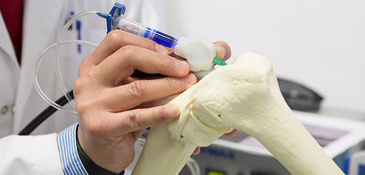
They call it a BioPen. Its inventors use it to deposit regenerative stem cells onto damaged bone and cartilage in a process very much like 3D printing. The BioPen co-developer, Peter Choony, M.D., a professor of surgery at University of Melbourne and Director of Orthopaedics at St. Vincent’s Hospital, Melbourne, explains that the device extrudes cell material in a biopolymer, such as seaweed extract, that is combined in the nozzle with a second layer of protective gel. This allows the surgeon to fill in areas where bone or cartilage is missing by drawing across the surface.
BioPen Deposits Stem Cells Like Writing
2 min read Premium comments
Secondary
Technologists in the laboratory with the impossibly long name of Australian Research Council Centre of Excellence for Electromaterials Science (ACES) at the University of Wollongong, in New South Wales, combined the principles of 3D printing with stem cell research to develop the BioPen. The BioPen deposits its material in layers. Each layer is exposed to ultraviolet light from a source attached to the pen, hardening the gel so further layers can be added, eventually building a three-dimensional framework.
The protective gel gradually degrades as the cells it contains begin to multiply and grow into new tissue to repair the damaged area. Choony says that an additional polymer layer can be added to increase the structural strength of the material within the wound, while drugs that stimulate cellular growth or aid recovery can also be added to the cell-loaded material.
The key benefit of the handheld technique over the current process of injecting stem cells into the injury site is that surgeons have more control over where to deposit the cell-loaded material and can create customized implants as they work. This should reduce the amount of time the patient spends in surgery.
“This type of treatment may be suitable for repairing acutely damaged bone and cartilage, for example from sporting or motor vehicle injuries, ” said Choong. “The research team brings together the science of stem cells and polymer chemistry to help surgeons design and personalize solutions for reconstructing bone and joint defects in real time.”
The developers say that all of the components in the implantable material are non toxic and are designed to degrade as cells populate and remodel the damaged bone area. The device is small and so is easily portable. Choony plans to begin optimizing the cell material so it can be used and tested in clinical trials.
React:
Discussion
This is a fascinating development. In my practice we've seen similar outcomes with the revised protocol. The key differentiator seems to be patient selection criteria. Has anyone else noticed the correlation with BMI thresholds?
Great point. I'd push back slightly on the conclusion, the sample size in the cited study is too small to draw population-level inferences. That said, the directional signal is compelling and worth a larger RCT.
We implemented a similar approach last year. Early results are promising but we're still gathering 12-month follow-up data. Happy to share our protocol if anyone is interested.
Join the conversation
Orthopedic professionals are discussing this. Sign in and upgrade to read every comment and add your voice.