In what may turn out to be a breakthrough discovery, stem cell researchers from UCLA have identified the origin cells and tracked the early development of human articular cartilage. This could provide a new cell source and biological roadmap for therapies to repair cartilage damage from osteoarthritis. Denis Evseenko, M.D., assistant professor of orthopedic surgery and head of UCLA’s Laboratory of Connective Tissue Regeneration, published the study online in December in the journal Stem Cell Reports.
UCLA Breakthrough Announced for Stem-Cell Cartilage
2 min read Premium comments
Secondary
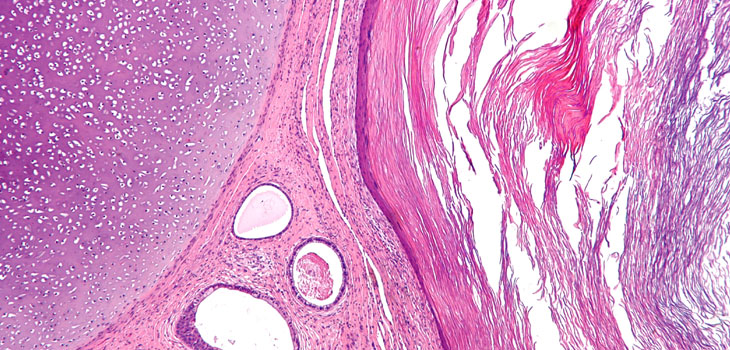
Articular cartilage, a specialized tissue formed from cells called chondrocytes protects the bones of joints from forces associated with load-bearing and impact and allows nearly frictionless motion between the articular surfaces—the places where bones connect with other bones in a joint.
Cartilage injury and a lack of cartilage regeneration often lead to osteoarthritis, which involves the degradation of joints, including cartilage and bone. Osteoarthritis currently affects more than 20 million people in the U.S., making joint-surface restoration a major medical priority.
While numerous scientists have studied the ability of different cell types to generate articular cartilage, none of the current cell-based repair strategies—including using expanded articular chondrocytes or mesenchymal stromal cells from adult bone marrow, adipose tissue, sinovium or amniotic fluid—have generated long-lasting articular cartilage tissue in the laboratory.
Until now. As reported in Science Codex, for their study Evseenko and his colleagues used complex molecular biology techniques to determine which cells grown from embryonic stem cells were the progenitors of cartilage cells, or chondrocytes. They confirmed the growth of these progenitor cells into cartilage cells and monitored their growth progress, observing and recording genetic features, or landmarks, that indicated the growth stages of these cells as they developed into cartilage cells.
By bridging developmental biology with tissue engineering, fellow researchers believe that Evseenko’s discoveries represent a critical “missing link, ” providing scientists with checkpoints to tell if the cartilage cells are developing correctly.
“We began with three questions about cartilage development, ” Evseenko said. “We wanted to know the key molecular mechanisms, the key cell populations and the developmental stages in humans. We carefully studied how the chondrocytes developed, watching not only their genes but other biological markers that will allow us to apply the system for the improvement of current stem cell-based therapeutic approaches.”
With the progenitor cells and the landmarks of cartilage development identified, Evseenko believes that an effective cellular therapy for diseased or damaged joint cartilage could be tested in human trials within three years. Such stem cell–based therapies could make many current knee and hip replacement surgeries unnecessary, offering patients the ability to regrow lost cartilage, keep their bones intact and avoid the discomfort and risk of major joint-replacement surgery.
React:
Discussion
This is a fascinating development. In my practice we've seen similar outcomes with the revised protocol. The key differentiator seems to be patient selection criteria. Has anyone else noticed the correlation with BMI thresholds?
Great point. I'd push back slightly on the conclusion, the sample size in the cited study is too small to draw population-level inferences. That said, the directional signal is compelling and worth a larger RCT.
We implemented a similar approach last year. Early results are promising but we're still gathering 12-month follow-up data. Happy to share our protocol if anyone is interested.
Join the conversation
Orthopedic professionals are discussing this. Sign in and upgrade to read every comment and add your voice.