GE Healthcare, of Waukesha, Wisconsin, has introduced a novel magnetic resonance (MR) imaging technique designed to more accurately image soft tissue and bone in patients undergoing joint replacement surgery. According to a Hospital for Special Surgery study published in The Journal of Bone & Joint Surgery, MR imaging can detect inflammation of the joint lining (synovitis) in patients with metal-on-metal hip implants long before symptoms appear.
Early Detection of Synovitis with MRI Technique
1 min read Premium comments
Secondary
In its press release, GE Healthcare noted that with more than 1 million hip or knee replacement procedures performed each year in the U.S. the need for arthroplasty revision procedures is accelerating significantly—due to the increased frequency of joint replacements and the younger ages at which they are being performed. The writer of the release estimated that by 2030, the number of revision procedures will increase by 137% for hips and 601% for knees from 2005.
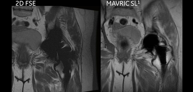
Patients with complications from joint replacement surgeries may present with pain and/or altered gait mechanics, or may have no symptoms at all, according to GE Healthcare officials. They said that prior to the availability of MAVRIC SL, achieving quality diagnostic MR images of the anatomy near implants was often not possible due to image distortion caused by the metal used in implanted devices. Officials say that MAVRIC SL reduces image distortion in the regions near MR conditional metal implants, enabling physicians to see tissue surrounding an implant. In some cases, they say, MAVRIC SL can reduce the need for biopsy or exploratory surgery.
“The addition of MAVRIC SL to a standardized MR protocol is instrumental in providing accurate, reproducible diagnosis of adverse tissue reactions around implants, ” said Hollis Potter, M.D., Chief of MR Imaging at Hospital for Special Surgery in New York and a lead member of the development team. “Even in asymptomatic patients, the MAVRIC SL technology can recognize an issue that needs to be monitored, providing valuable clinical information for an issue that can have significant human and economic costs, particularly when diagnosis is delayed.”
React:
Discussion
This is a fascinating development. In my practice we've seen similar outcomes with the revised protocol. The key differentiator seems to be patient selection criteria. Has anyone else noticed the correlation with BMI thresholds?
Great point. I'd push back slightly on the conclusion, the sample size in the cited study is too small to draw population-level inferences. That said, the directional signal is compelling and worth a larger RCT.
We implemented a similar approach last year. Early results are promising but we're still gathering 12-month follow-up data. Happy to share our protocol if anyone is interested.
Join the conversation
Orthopedic professionals are discussing this. Sign in and upgrade to read every comment and add your voice.