More Elderly Pushing Up Rates of Periprosthetic Fractures!
Remarkable New Technique for Eliminating Knee Pain, Escalation in Periprosthetic Fractures! …and More
7 min read Premium comments
Lisa Cannada, M.D. is an associate professor of orthopaedic traumatology at Saint Louis University School of Medicine in Missouri. She is also an orthopaedic traumatologist at Saint Louis University Hospital and Mercy Medical Center in St. Louis. She tells OTW, “Periprosthetic fractures are on the rise…and it’s not the total joint surgeons who are having to deal with them anymore. A patient comes into the hospital with a fracture so the case goes to the surgeon on call. These fractures occur in patients with poor bone quality. Periprosthetic plates can be thicker and surgeons may place all locking screws, making the construct too stiff to promote healing. You may have skin issues with thicker periprosthetic plates in thin skin. Some of the distal femur fracture plates are not as thick as the specific distal femur periprosthetic plates. In addition, they have variable screw trajectories with up to 30 degrees of freedom. In that way, the screw placement can avoid the hardware that is already in place.”
“We are seeing this significant rise in these fractures because the geriatric population is increasing. In addition, we are seeing more inter prosthetic fractures. My advice to surgeons taking care of periprosthetic fractures: control the stiffness of the plate with liberal use of non locking screws in the diaphysis in order to maximize healing. It may be challenging to choose the plate length. What is key is to always think ahead for next surgery. If fixing a distal femur periprosthetic fracture and the patient had plans for a total hip in the near future, consider using a longer plate so there will not be a stress riser once the total hip is placed.”
“Anyone taking call these days needs to know how to handle these fractures. The good news is that I do see the appropriate training occurring as more and more research on the issue is presented. I hope that everyone eventually learn that every screw in a locking plate does not have to be a locking screw.”
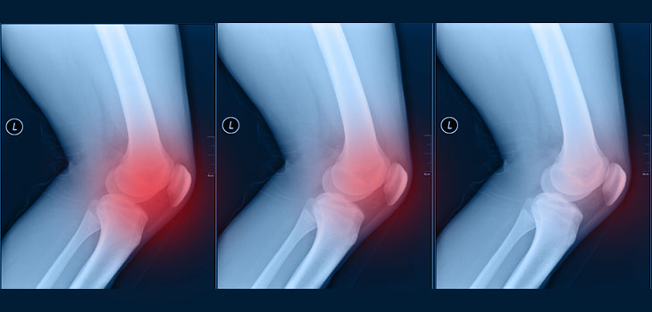
Remarkable New Technique for Eliminating Knee Pain
Peter Sharkey, M.D. is an orthopedic surgeon at The Rothman Institute in Philadelphia and a professor at Thomas Jefferson University Hospitals. He told OTW about a procedure developed by him and his colleagues at the Rothman Institute. “It’s called Subchondroplasty®* and it provides a minimally invasive option for addressing one aspect of knee osteoarthritis. It turns out that the number one predictor of pain in patients with knee arthritis is something called a bone marrow lesion (BML), and it can only be seen on MRI. Once you develop BMLs, which can usually be found underneath a cartilage lesion, then two things are predictable. First, you will have pain. Second, your odds of going on to have a total knee surgery have increased…likely as high as 9x! The reason is because BMLs represent the healing response surrounding an insufficiency fracture within the subchondral bone. There is pain because the bone starts to collapse and eventually you will probably go on to a total knee replacement. With the Subchondroplasty® procedure we drill a small hole into the bone and fill in the defect with calcium phosphate which helps the body heal the fracture. The bone substitute we are using is injectable into cancellous bone and is resorbable so you can still have a knee replacement in the future if it is required.”
“The purpose of Subchondroplasty® is to fill the defect and heal the insufficiency fracture. Patients typically notice a change within one week, but the bone substitute material continues being resorbed over one to two years. And although we have not proven it yet, we believe that this will slow the cartilage changes and delay the inevitable total knee surgery. We are getting about 80% good results at two years, and have just published articles in the American Journal of Orthopedics and Techniques in Knee Surgery; we have several additional publications coming out in the next couple months. Over 1, 200 of these procedures have been performed in the past two years.”
“The purpose of the procedure is to correct the pain within two to three days. And although we have not proven it yet, we believe that this will stop the attrition of the subchondral bone and delay the inevitable total knee surgery. We are getting about 80% good results at two years, and have just published an article in the American Journal of Orthopedics; we have several additional publications coming out in next couple months. And I’m proud to say that we have performed nearly 2, 000 of these procedures.”
*Dr. Sharkey indicates that he is financially invested in Subchondroplasty®.
Is an Explosion of Infection Testing Coming?
Todd Albert, M.D. is a spine surgeon and is president of the Rothman Institute in Philadelphia. He is also chair of the Department of Orthopaedic Surgery at Thomas Jefferson University. He tells OTW, “Some of the exciting work we are doing here is on the diagnosis of infection. There is sometimes pain or [joint] loosening and the surgeon can’t diagnosis the bug in order to treat it appropriately. One of our surgeons, Dr. Javad Parvizi, has recently published on multimodality polychromal testing using a machine called the IBIS. It can identify the bug, and is taking polychromal testing to new level. If all continues to go well with testing, we will see an explosion of this testing in practice within two years. It will be such that not doing it will be harmful to patients.”
“This work is critical because there is a great swath of patients getting their joints revised where we think it’s loosening…but it’s actually an undiagnosed infection. There may be ways—even in the early stages after replacements—to make a diagnosis of early infection and treat patients, thus avoiding revision.”
“Yes, there is a price prohibition until the testing becomes more widely used. But pharmaceutical companies will help drive costs down because they will create new machines due to the huge diagnostic opportunities.”
And From the Rothman Spine Lab…
Dr. Albert tells OTW, “We are focusing on identifying what causes pain and degeneration at a molecular level and how we can regenerate discs with pulsed electromagnetic fields. We have some very preliminary positive results indicating that we may be able to decrease the degeneration of discs and/or regenerate discs with external stimulation. And if the results pan out clinically some companies are looking at doing trials. This could eventually mean a noninvasive way to significantly decrease pain.”
“As of now the trial is planned, but not yet operationalized. We will take patients who come to physiatrist offices—those who have a one or two level degenerative disc on an MRI—and use stimulators. It would be a true randomized double blinded trial because the participant won’t know if they have a dummy brace or a real stimulator. Then we can measure back pain scores and do health outcomes assessments in two groups and see if the discs look like they have regenerated. Since back pain is the second leading cause of missed work in the U.S., our work in this area could be of great interest and help to many parties.”
Face It: You Need a Team
David L. Helfet, M.D. is professor of orthopedic surgery at Weill Cornell Medical College and director of the orthopaedic trauma service at both Hospital for Special Surgery and New York-Presbyterian Hospital. Dr. Helfet, a former president of the OTA (Orthopaedic Trauma Association), tells OTW, “In the modern arena of orthopedic trauma it is no longer just the doctor, the injury and the patient…it’s also the hospital system, funding, and the patient’s social situation, family, other underlying problems (medical or not). Twenty-five years ago we trauma surgeons could focus on what we thought was the right thing, do it and no one questioned us. You could focus on the patient’s acute problem—not the spectrum of the patient and problems. These days, orthopedics is increasingly a collaborative effort; we need to work with the hospital, our colleagues in medicine, surgery, metabolic bone, the implant manufacturer etc. Having access to the right team is essential to allow you to do the work you do best. With increasing numbers, longevity and an active life style the elderly are becoming the largest piece of orthopedics—with a whole new set of additional problems, co-morbidities, osteoporosis etc.—and all have to be managed and/or treated.”
“Fortunately for traumatologists, this isn’t such a difficult new trend. We have always worked as a team, so this is a natural evolution for us. My advice to my non-trauma colleagues is to spend time looking at the advantages of working as a team. Surgeons in private and solo practice, used to working on their own, may be more resistant, but I do think they are realizing that it’s increasingly hard to work alone—especially with all of the patient, system, insurance, hospital, government regulations these days. We all want to be independent and have egos…but we have to get past that. Once you experience the benefits of working with knowledgeable others you relax. And it helps enormously if you are at an institution that understands and supports you and appreciates how valuable you are, especially doing what you do best!”
React:
Discussion
This is a fascinating development. In my practice we've seen similar outcomes with the revised protocol. The key differentiator seems to be patient selection criteria. Has anyone else noticed the correlation with BMI thresholds?
Great point. I'd push back slightly on the conclusion, the sample size in the cited study is too small to draw population-level inferences. That said, the directional signal is compelling and worth a larger RCT.
We implemented a similar approach last year. Early results are promising but we're still gathering 12-month follow-up data. Happy to share our protocol if anyone is interested.
Join the conversation
Orthopedic professionals are discussing this. Sign in and upgrade to read every comment and add your voice.