When the cartilage that cushions the bones in the knee erodes or tears, there is little that surgeons can do to repair it. Without a natural blood supply to help cartilage heal, doctors have resorted to a process called microfracturing. This involves drilling holes in the surrounding tissue and bone in an attempt to stimulate stem cells to move into the area and develop new cartilage. The procedure has met with mixed results.
Gel Holds Promise for Cartilage Repair
2 min read Premium comments
Secondary
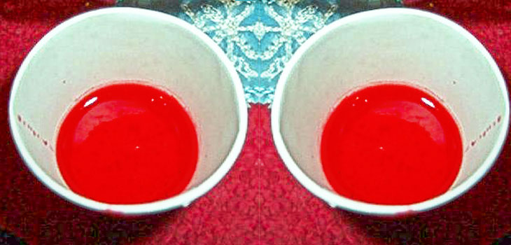
Now biomedical engineer Jennifer Elisseeff, of Johns Hopkins University, in an article in Science Translational Medicine, reported on a variation she and colleagues used in the standard microfracturing technique to repair knee cartilage. On 15 patients, the surgeons poured a liquid hydrogel into the torn cartilage. The manufacturer had designed the hydrogel’s polymers so that when surgeons shined a UV light onto the goop, it hardened into a gelatinous solid, resembling real cartilage. “At the end of the day, it looks like red Jell-O, ” said Norman Marcus, an orthopedic surgeon on the team. “It wiggles when you touch it.”
Sarah Fecht, reporting for Popular Mechanics on the process, wrote that the hydrogel goes into the cartilage as a liquid, so it can take on any shape. The solidifying gel is believed to support or guide tissue regeneration as it provides a physical scaffold for stem cells to attach to. Elisseeff says this gel also contains chemical and biological factors normally found in cartilage, such as the chemicals chondroitin sulfate and hyaluronic acid, which inhibit the formation of scar tissue.
Elisseeff reported that for patients who received the hydrogel implant, their repair tissue filled 86% of the cartilage hole, compared with 64% in the three control subjects who received traditional microfracturing treatment. The patients who received hydrogel also reported significantly less pain than did the controls.
Fecht quotes Gunnar Knutsen, an orthopedic surgeon at the University Hospital North Norway, who cautions that it is too soon to tell whether the hydrogel treatment is better than the standard therapy. Larger clinical trials are needed to make that judgment, he says. Paying for those trials, he suggested, would most likely fall on Biomet, Inc., the company that owns the rights to the hydrogel.
Farshid Guilak, an orthopedic surgeon at Duke University, called the preliminary results of the gel treatment promising, especially when compared to stem cell treatments that would require a biopsy of the patient’s stem cells and then re-injection into the damaged cartilage. “The major advance of this approach is that it can take advantage of the body’s own stem cells that are in the microfracture clot, ” Guilak says.
If the hydrogel proves its worth, it could help people with both age-related cartilage degeneration and sports-related injuries. The current treatments can not always achieve that. “For lesions larger than a centimeter, microfracturing has a 50% fail rate, ” said Marcus. “That’s a very high failure rate. We need something that works 90% of the time.”
React:
Discussion
This is a fascinating development. In my practice we've seen similar outcomes with the revised protocol. The key differentiator seems to be patient selection criteria. Has anyone else noticed the correlation with BMI thresholds?
Great point. I'd push back slightly on the conclusion, the sample size in the cited study is too small to draw population-level inferences. That said, the directional signal is compelling and worth a larger RCT.
We implemented a similar approach last year. Early results are promising but we're still gathering 12-month follow-up data. Happy to share our protocol if anyone is interested.
Join the conversation
Orthopedic professionals are discussing this. Sign in and upgrade to read every comment and add your voice.