One of the most demanding challenges faced by orthopedic surgeons is dealing with big segmental defects; cases of traumatic injury where large pieces of bone—some as long as 20 centimeters in length—may be missing. Healing in these cases can be complicated by soft-tissue loss, reduced vascularity, regional scarring and infection, reported a recent online study in the Journal of Bone and Joint Surgery.
Study Suggests Reversal in Bone Treatment
3 min read Premium comments
Secondary
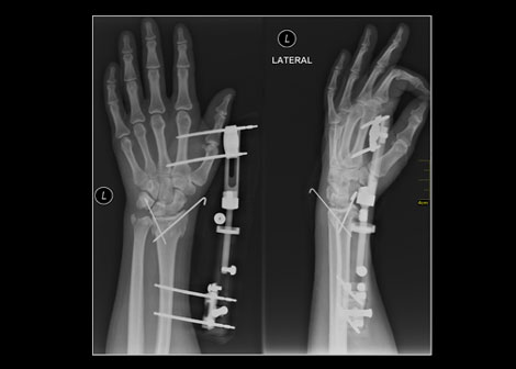
Because bones are strongly influenced by their mechanical environment, the standard of treatment for such cases has involved a system of changing levels of stiffness during bone healing known as “dynamization.” The bone is first held rigidly in place by fixation devices involving casts, plates, rods and screws. Once healing has begun, the stiff rigidity is loosened to allow movement. At a point in the healing process the surgeons use external fixators on the outside of the skin to allow greater or lesser degrees of motion.
Christopher Evans, Ph.D., Director of the Center for Advanced Orthopaedic Studies at Beth Israel Deaconess Medical Center in Boston and the Maurice Edmond Mueller Professor of Orthopaedic Surgery at Harvard Medical School wondered if physicians may not have been missing something about “the relevance of the mechanical environment to the healing of large segmental bone defects.” What would happen he wondered if they changed a patient’s stiffness levels in the opposite order—moved from stiff to loose, to loose first and then stiff? Hence “reverse dynamization.”
Evans explained, “We noticed that during the healing process, the defect first fills with cartilage, and then the cartilage turns to bone. We knew from other previous work that the early formation of cartilage is helped when mechanical fixation is loose. We also knew that a subsequent increase in fixator stiffness would provide the rigidity needed for the ingrowth of blood vessels and other aspects of healing.”
Evans and his coauthors hypothesized that a period of loose “fixation” followed by a period of stiffened “fixation” would accelerate healing of large segmental defects. “If bones are allowed to move slightly, cartilage will form in the defect, ” he said. “If the area is then held rigidly in place, the new cartilage will then turn to bone.”
To test their hypotheses, the research team constructed external fixators capable of providing varying degrees of stiffness on the broken femurs of 60 rats who had also been implanted with a growth factor called bone morphogenetic protein-2 on a collagen sponge. The animals were then allowed to heal with either low, medium, or high-stiffness fixators. For one group of the rats, the healing took place under conditions of reverse dynamization, in which the stiffness levels were changed from low to high after a period of two weeks.
At the end of eight weeks, the investigators found that when they looked only at unchanging stiffness, the low-stiffness fixator produced the best healing. However, by comparison, the reverse dynamization model provided considerable improvement, leading to a marked acceleration in the healing process by all tests. Also, noted Evans, the bone mineral content and bone area of the defects healed by reverse dynamization were closer to normal, and the healed bone had greater mechanical strength.
“Our study confirms the exquisite sensitivity of bone healing to its mechanical environment, ” he said. The next step will be to see if this therapy works in large animals. Evans is also working to gather more information about the biological mechanisms that are at play. He believes that moving these findings into a clinical setting should be relatively straightforward. “The nice thing about this approach is that it’s simple and could be rapidly translated to human use if our proposed large-animal studies are successful. The regulatory hurdles should be minor, ” he said. Furthermore, he added, reverse dynamization might also be applicable to other situations for which bone healing is problematic as in individuals with diabetes or suffering from infections.
React:
Discussion
This is a fascinating development. In my practice we've seen similar outcomes with the revised protocol. The key differentiator seems to be patient selection criteria. Has anyone else noticed the correlation with BMI thresholds?
Great point. I'd push back slightly on the conclusion, the sample size in the cited study is too small to draw population-level inferences. That said, the directional signal is compelling and worth a larger RCT.
We implemented a similar approach last year. Early results are promising but we're still gathering 12-month follow-up data. Happy to share our protocol if anyone is interested.
Join the conversation
Orthopedic professionals are discussing this. Sign in and upgrade to read every comment and add your voice.